12 Month Subscription
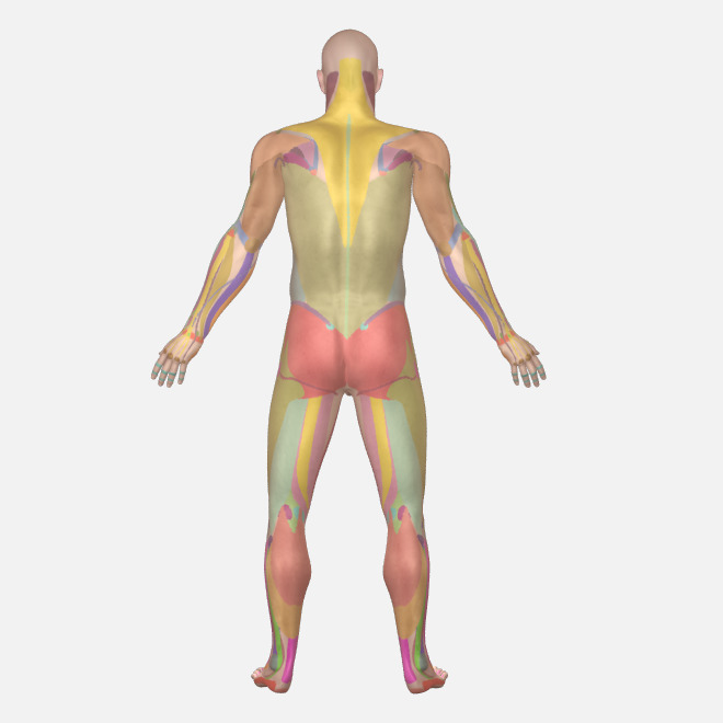
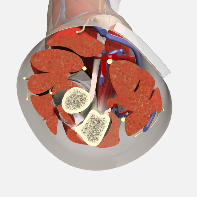
Urology
Primal’s interactive clinical solution for successful outcomes
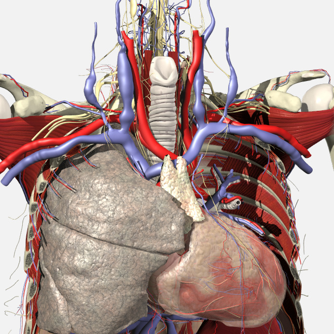
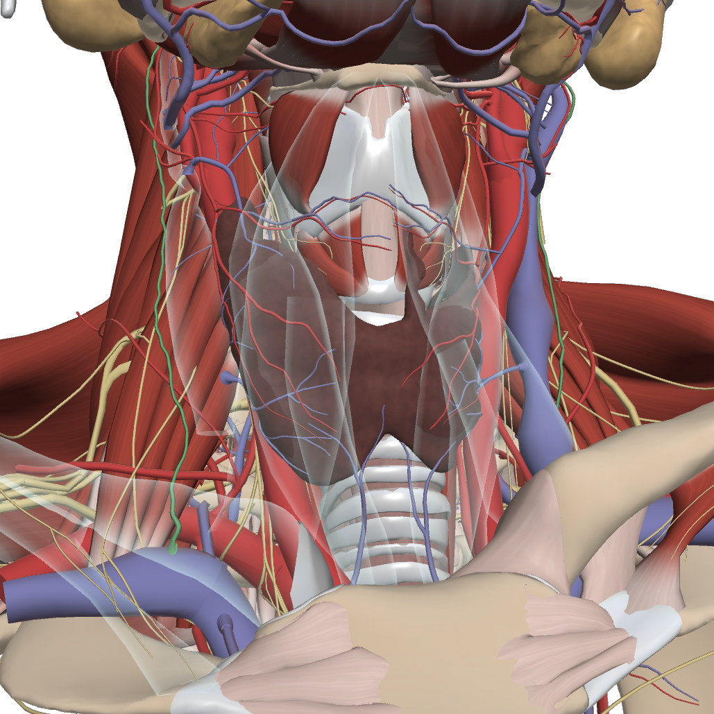
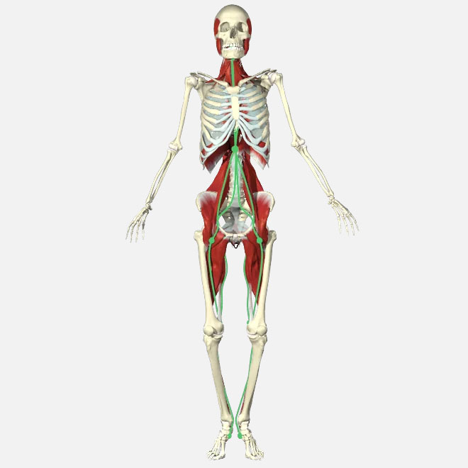
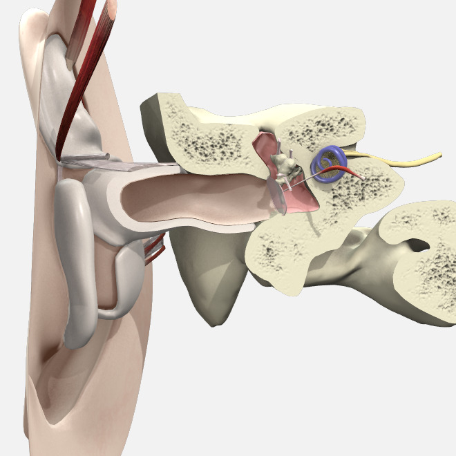
This career-spanning resource will be all you need to see you from student to practitioner. With over 50 interactive 3D anatomy models chosen for their focus on the urinary system from both male and female anatomy, review anatomy down to the smallest of structures. Hone your skills with real-life clinical movies and text articles written by professionals in the field and engage with patients to have the best outcomes possible with a library of patient education resources ready for you to use.