Aorta tell you how much I love you!
In this blog series, we take a deeper look into anatomical variations to celebrate our differences, as well as shine a spotlight on potential issues they may cause. We are all distinctive in the way we look, think and behave, and so is our anatomy. Yes, we have organs in common, but their shape and position can vary from individual to individual.
This article will look at the anatomy and function of the aorta, the different variations in the branching patterns of the aortic arch and how this could affect us.
It should be noted that these variations are so common that they are present in over one-third of some populations. This Irish Journal of Medical Science study and this Surgical and Radiologic Anatomy study confirm how critical this knowledge can be when assessing patients for thoracic, neck and thyroid surgery.
Anatomy of the aorta
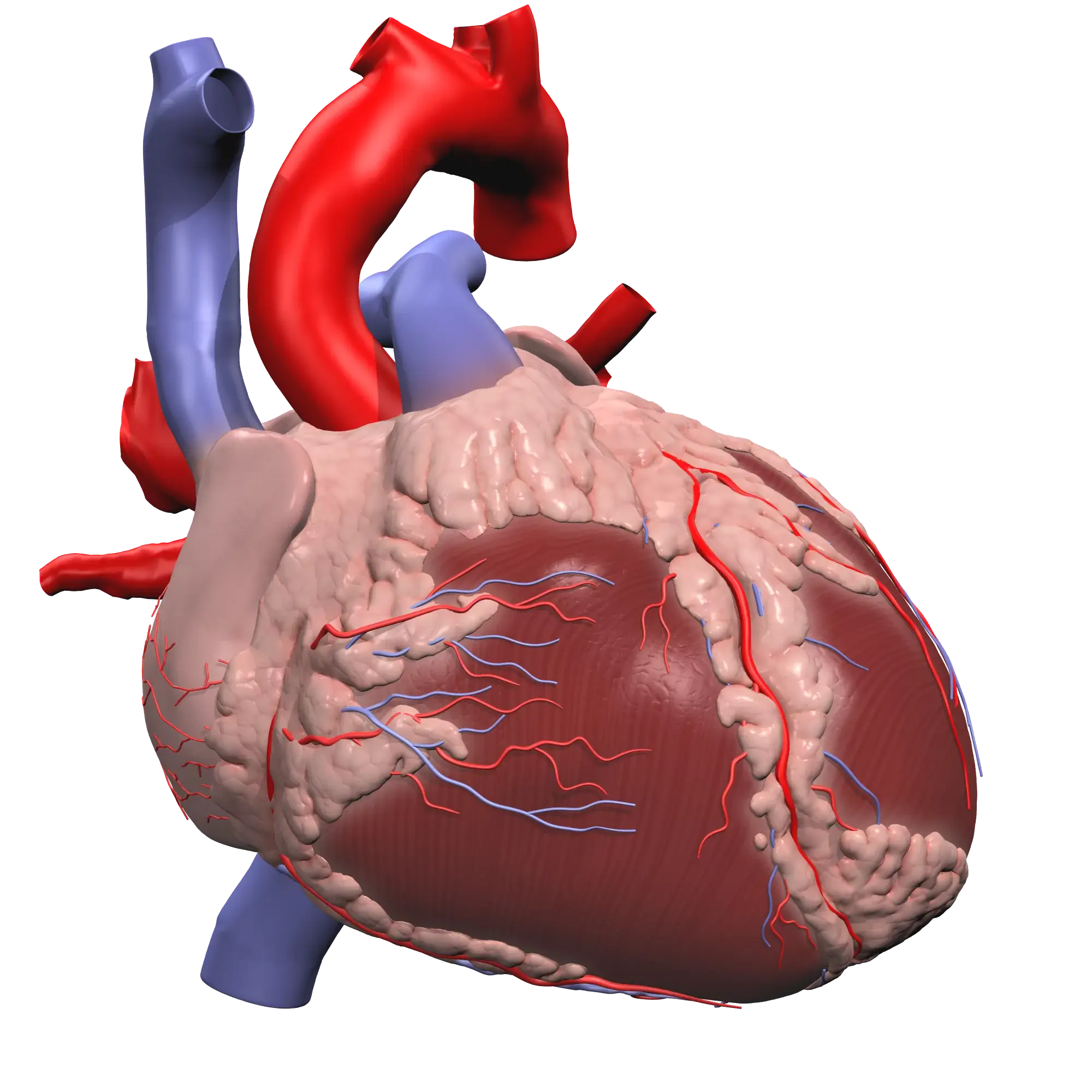
The aorta is the largest artery in the human body. It arises from the left ventricle of the heart, passes back over the heart, then down through the thorax and abdomen before dividing into the common iliac arteries and into the pelvis.
Due to its size, the aorta is described in four parts:
-
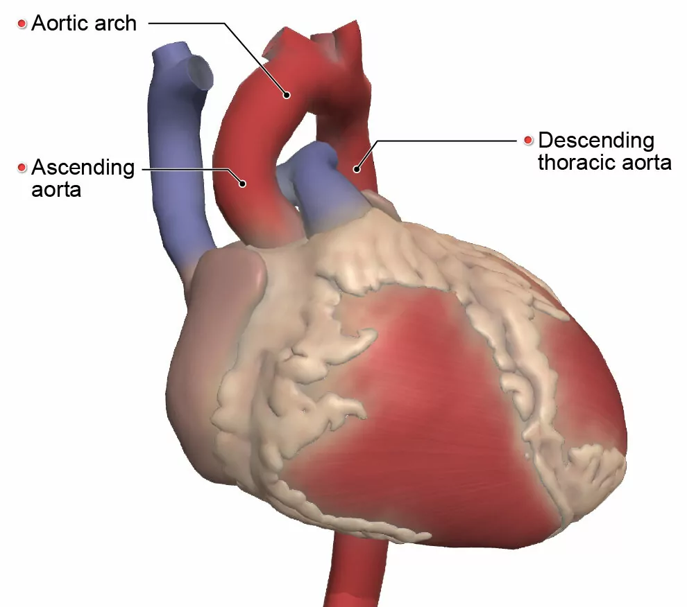
- The ascending aorta ascends approximately 5 cm and is next to the pulmonary trunk.
- The aortic arch arches posteriorly over the left main bronchus, which is a large airway to the left lung.
- The descending thoracic aorta begins at the level of the fourth thoracic vertebra and descends through the diaphragm to become the abdominal aorta.
- The abdominal aorta descends to the abdomen region and divides into the left and right common iliac arteries at the level of the fourth lumbar vertebra.
Anatomical variations in the branching patterns of the aortic arch
Before diving in, we should examine the anatomy of the aortic arch in more detail. The aortic arch is convex-shaped and is a continuation of the ascending aorta. It begins at the level where the second rib joins the sternum on the right side of the body, then passes over the trachea, crossing to its left side.
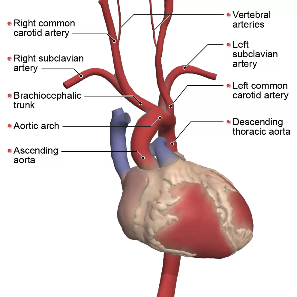
The aortic arch supplies fresh blood to the head, neck and upper limb regions via its branches. Usually (in 80% of cases according to this article), it has three main branches:
-
- The brachiocephalic trunk branches into the right common carotid artery and the right subclavian artery supplying the right side of the head, neck and right arm.
- The left common carotid artery branches at the carotid sinuses into the internal and external carotid arteries. This supplies the neck, face, skull, middle ear, brain, pituitary gland (hypophysis), orbit and choroid plexus.
- The left subclavian artery branches into the vertebral arteries, internal thoracic arteries, thyrocervical trunks, costocervical trunks and dorsal scapular arteries to supply the neck, thoracic wall, spinal cord, brain, meninges and upper limbs.
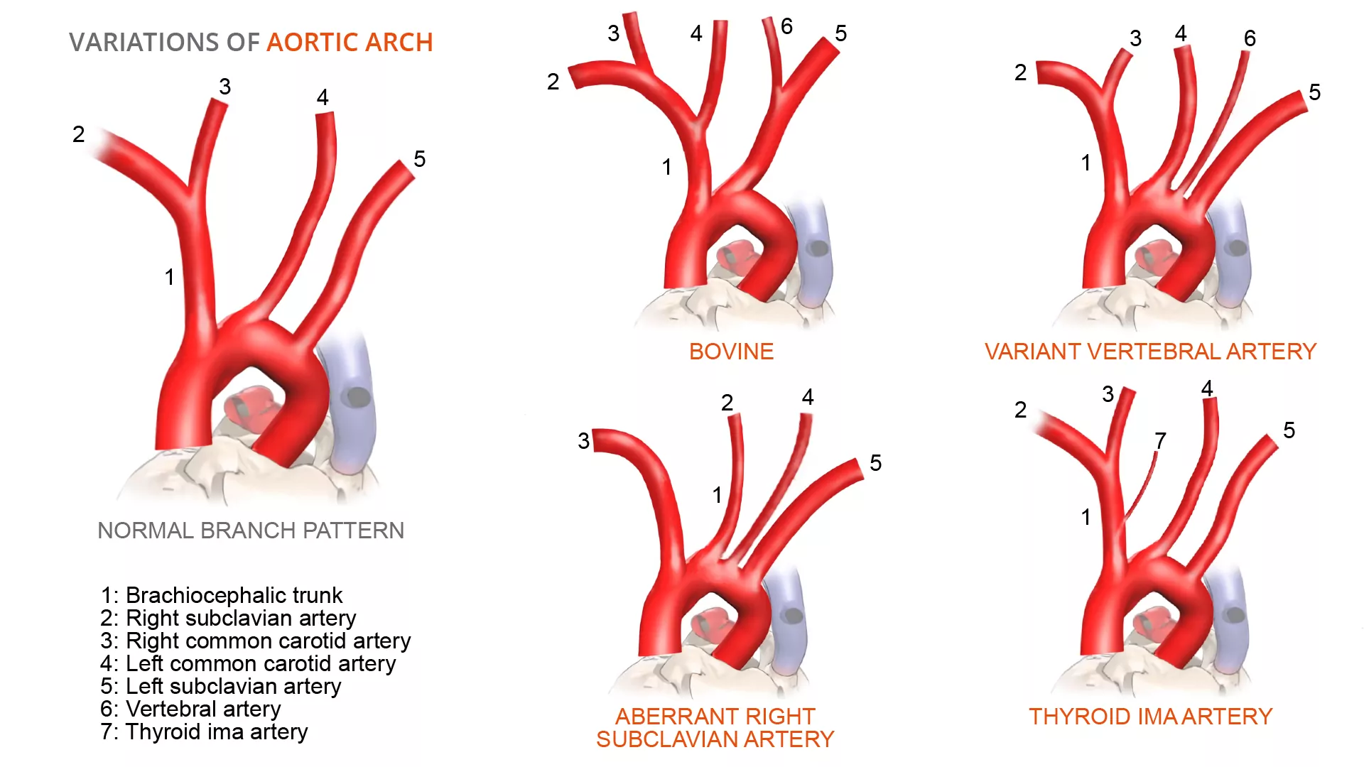
Although the above branching patterns are standard, sometimes they can differ, as outlined in these variations:
-
- Bovine arch – This occurs when the brachiocephalic artery and left common carotid artery share a common origin, while the right subclavian artery is present distal to this. This type of variation is present in approximately 15% of the population.
- Variant origin of vertebral arteries – Usually the vertebral arteries originate from the posterior superior portion of the subclavian artery on both sides to supply blood to the brain. However, in approximately 6% of cases they could rise directly from the aortic arch.
- Aberrant right subclavian artery – Instead of being a part of the brachiocephalic trunk, it arises as a separate branch directly from the aortic arch. This type of variation is present in approximately 0.5-2% of the population.
- Thyroid ima artery – This is an extra artery supplying the thyroid gland, apart from its main blood supply (superior and inferior thyroid arteries), that can arise from various locations within the branches of the aortic arch. This type of variation can be present in up to 15% of the population.
Why are there variations in the branching patterns of the aortic arch?
The development of the aorta is a complex process that begins during the third week of gestation. It involves the division and fusion of various pre-developed vessels, and anomalies can occur when these fail to form properly as outlined in this study. The variations can include differences in the distance between the origins of different branches or the number of branches that arise from the aortic arch as referenced in this article.
Some chromosomal abnormalities can potentially lead to these differences. It has been shown that there is a strong correlation (~98%) between patients with a bovine aortic arch and at least one congenital cardiac defect. And people with the aberrant right subclavian artery are 35% more likely to have Down syndrome.
Complications with variations in the branching patterns of the aortic arch
Although these variations are usually asymptomatic, they need to be identified as they can cause complications such as shortness of breath (dyspnea), difficulty with swallowing (dysphagia) and/or muscle pain as a result of a decrease in blood circulation. They can also cause complications during thorax and neck surgery, and can lead to misinterpretation in radiology exams.
Lastly, it is important to be aware of these variations as they can be a marker for thoracic aortic disease. Even if these variations do not lead to complications, they need to be clinically recognized as being relevant during surgical procedures. Ignoring them could lead to fatal consequences.
The images used in this piece have been taken from Primal’s 3D Atlas. To learn more about this or other Primal learning resources – please fill in the form here and our team will be in touch.