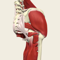
Osteopath, Daryl Herbert has been using Primal Pictures software since the first interactive skeleton was launched some years ago. When the first edition of the Interactive Anatomy Series was brought out, he quickly adopted it.
The material has been produced in such a way that it is easy to export images into presentations and I use this facility extensively when I am teaching.
When I first saw the Interactive Anatomy software I was extremely impressed," comments Daryl. "I had already been using the interactive skeleton for a couple of years but this new series was more realistic; giving me an almost living view of the joints, arteries, veins, muscles and ligaments, as well as the spine. It also included x-rays and MRI scans, pictures, slides and video clips demonstrating the bio-mechanics of movement.
“In fact this computerised, digital model of the spine that allowed me to view in 3D and rotate the images through 360 degrees, gave me a brand new way of teaching anatomy,” he explains. “Previously, anatomy had been taught to osteopathic students predominantly through two-dimensional images in text books – particularly if you didn’t have access to cadavers.
“Anatomy is one of the most important things that osteopaths have to learn. Although the actual information remains constant, there is a vast amount of knowledge that has to be acquired over a relatively short period of time. We have to know and understand all the internal, skeletal, vascular and neurological structures of the body and even if students are able to watch a dissection once a week, having the software enables us to supplement their learning and reinforce what they have seen in the cadaver lab.
“In some ways, students see more through looking at the software than they do by dissecting a cadaver. The software shows tissue in its ‘true’ state – ie. as it should be when they treat a patient — and provides dissection views so that they can see how the tissue is different in a cadaver and relate the two to put structures into the correct context.
“When I teach first year students basic osteopathic techniques, the software allows me to show them the bones, muscles and joints so that they can form an image in their heads of what I am explaining. I project images onto a large screen so that whilst I lecture, I can demonstrate the points I am making and relate them directly to the parts of the body in question. This gives them a much better feel for what they are supposed to be doing and many of them then access the software through the online resource to study and revise.
At a postgraduate level, I am able to use the spine to show diagnosis and pathology and then relate it to manipulation techniques. The material has been produced in such a way that it is easy to export images into presentations and I use this facility extensively when I am teaching. I know a lot of lecturers who use the software now in their teaching and we all find it immensely helpful. Once seen, Primal Pictures is the sort of product that people will adopt and use for themselves," he comments.
Daryl also finds the software extremely useful for patient education: “I often use the software when a patient doesn’t understand where their problem is and what is wrong,” he explains. “The images really help them to visualize what I am explaining. So, if their joints are not moving correctly, I can show them the position and orientation and explain what has happened and how I am going to treat it. This relaxes them, instills trust and confidence and aids greatly with patient compliance.”
